Medical Image Processing & AI
Advanced Artificial Intelligence and Computational Methods for Medical Diagnostics
Project Overview
In this project, we focused on applying advanced artificial intelligence and computational methods to solve complex problems in medical diagnostics and biomedical engineering. We utilized various machine learning models, particularly Convolutional Neural Networks (CNNs), to analyze diverse medical imagery. Our research demonstrated the use of AI for diagnosing COVID-19 from lung X-rays, segmenting brain lesions and breast tumors from MRIs and ultrasound, and diagnosing biomechanical conditions like fatigue foot. We also explored GEP for gastric cancer analysis and compared advanced models like SAM and U-Net, consistently aiming to improve diagnostic accuracy through computational intelligence.
Research Timeline & Achievements (2020–2025)
Across several studies (2020–2025), we applied data‑driven techniques to medical diagnosis. We used gene‑expression programming and sensitivity analysis to model how factors such as age, sex and H. pylori infection affect gastric cancer tumour location and size. During the COVID‑19 pandemic we developed CNNs to classify lung X‑ray images, achieving 93.2% accuracy, 96.1% sensitivity and segmenting infected tissue with 83.8% accuracy. Other projects introduced a quantum‑matched‑filter deep spiking neural network for MRI tumour segmentation, diagnosed foot fatigue using CNNs, combined robust PCA and CNNs to localise brain lesions, and compared U‑Net and Segment Anything models for breast tumour delineation.
Deep Learning
CNN & Neural Networks
Medical Imaging
X-ray, MRI & Ultrasound
Disease Diagnosis
COVID-19, Cancer & More
Research Visualization
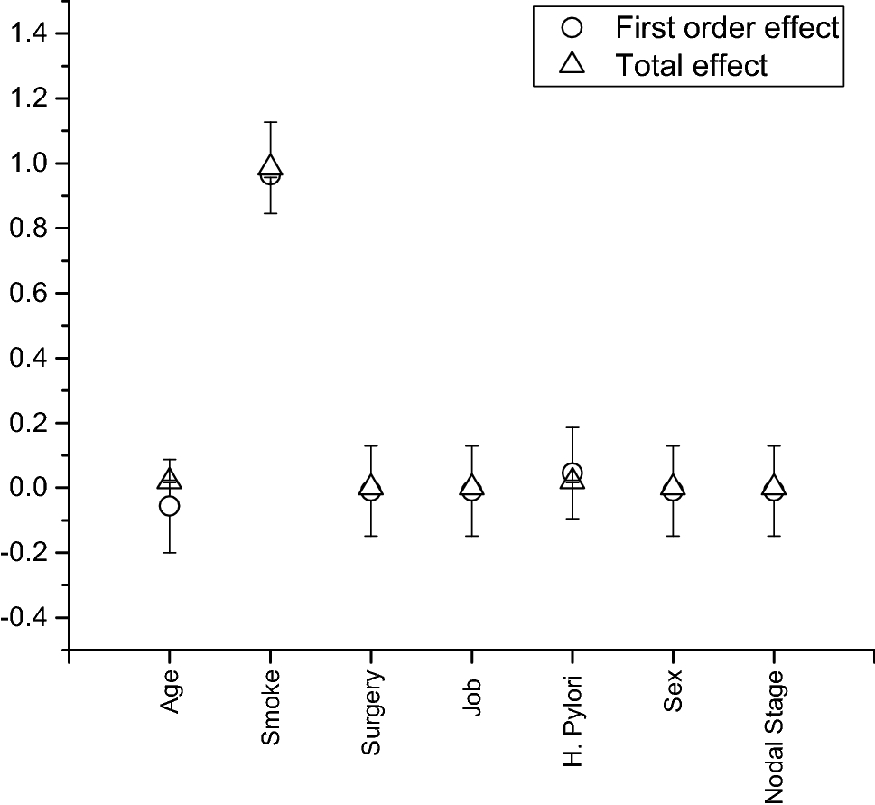
Gastric Cancer Analysis using Gene Expression Programming
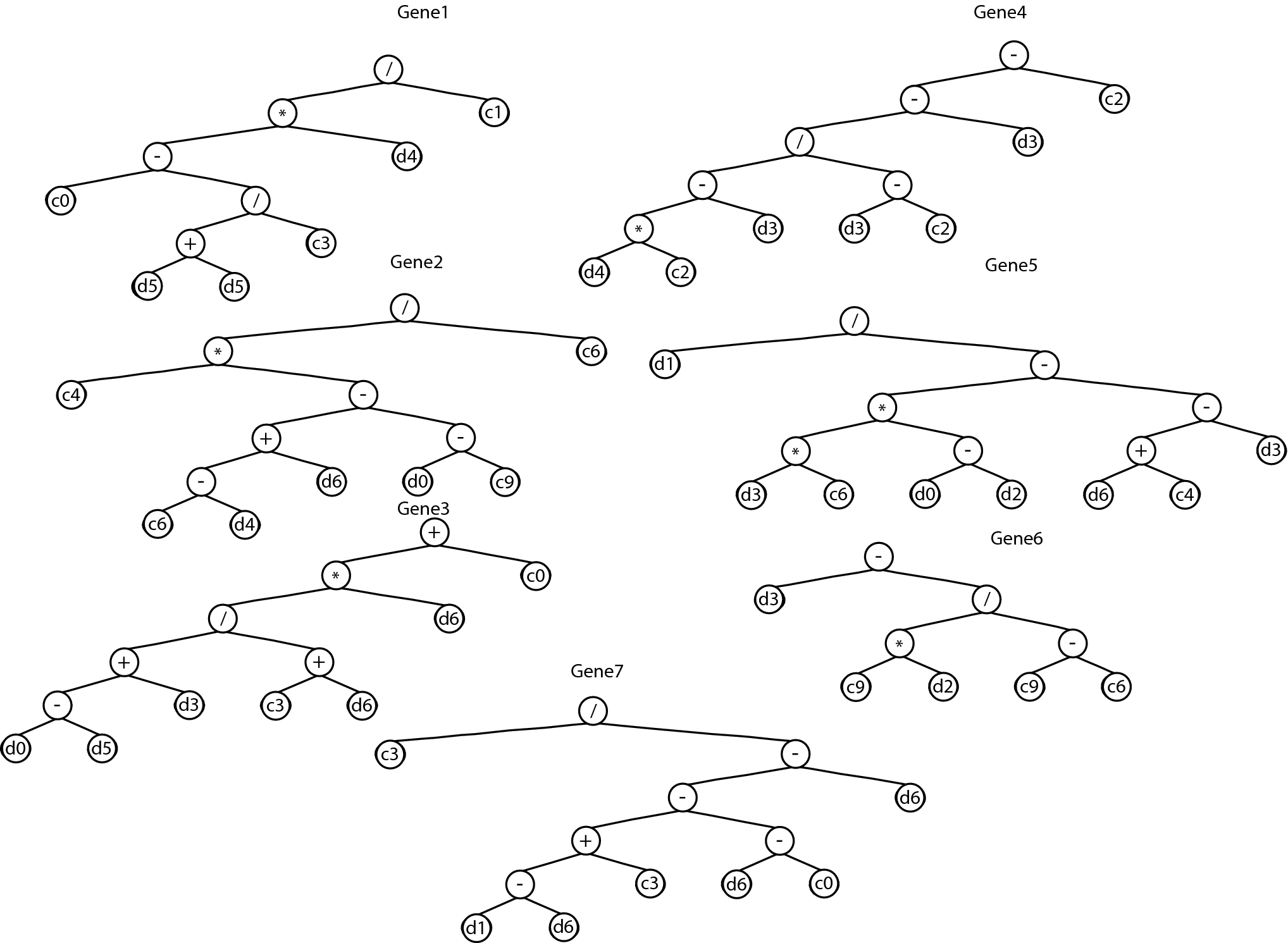
Gene Expression Programming Framework
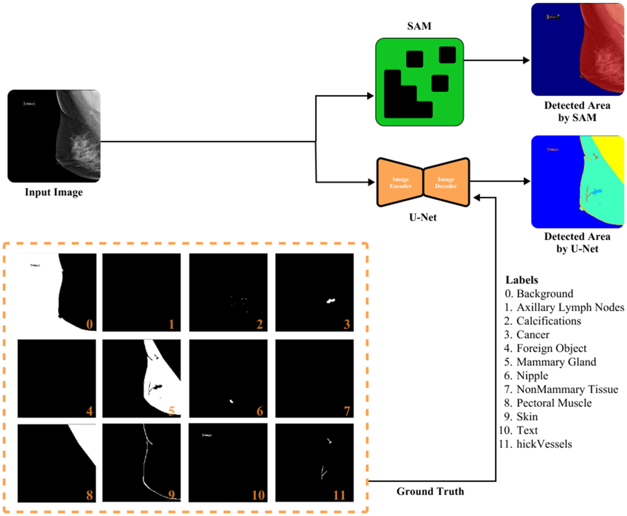
Medical Image Segmentation Results
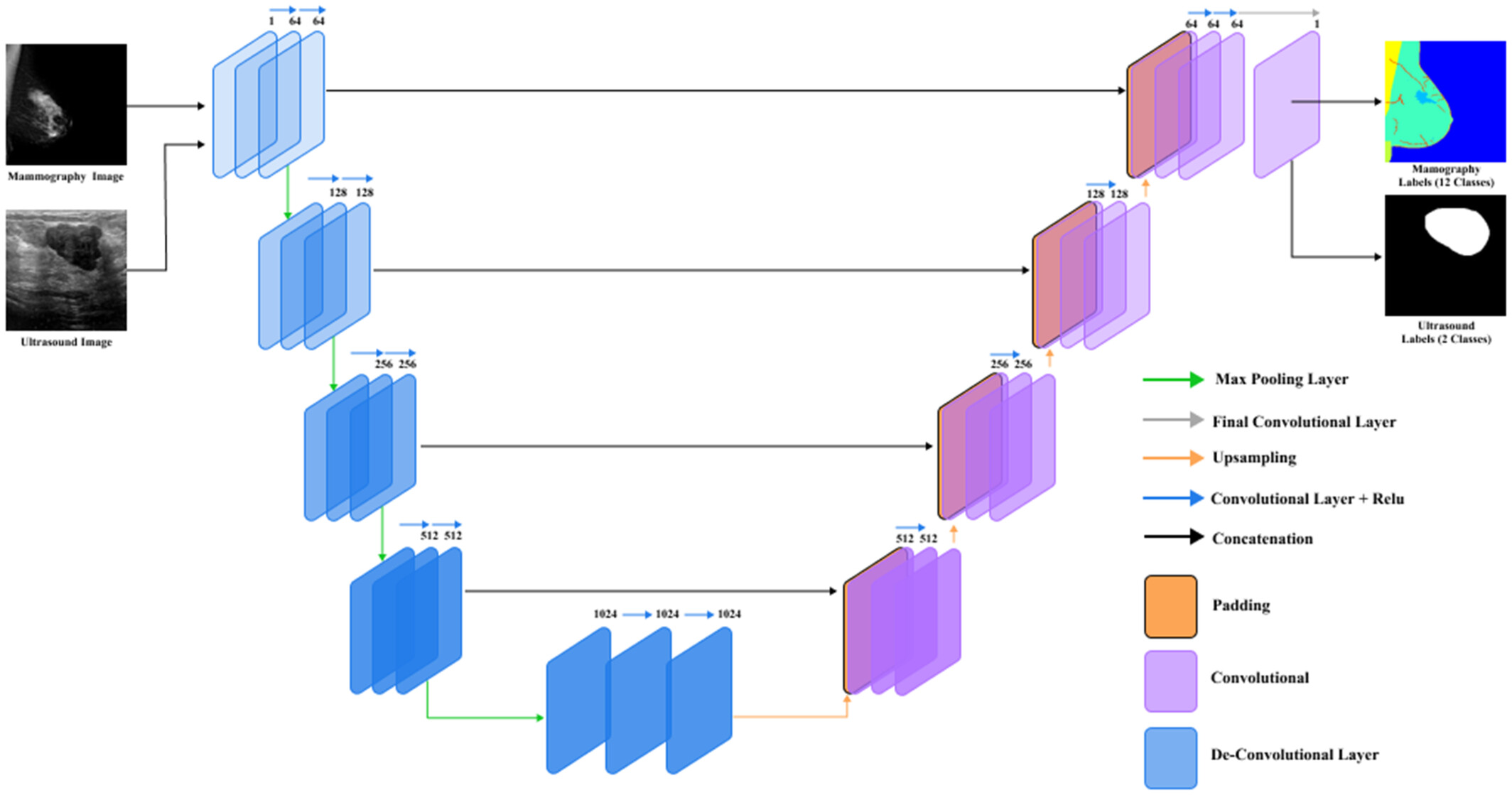
U-Net vs SAM Model Comparison for Breast Tumor Detection
Key Research Areas
COVID-19 Diagnosis
Detection and diagnosis of infected tissue in COVID-19 patients based on lung X-ray images using advanced convolutional neural network approaches.
Brain Lesion Detection
Detection of brain lesion location in MRI images using convolutional neural networks and robust Principal Component Analysis (PCA) techniques.
Breast Tumor Detection
Comparative analysis of Segment Anything Model (SAM) and U-Net for breast tumor detection in ultrasound and mammography images with improved accuracy.
Cancer Analysis
Application of gene expression programming for analyzing effective parameters in gastric cancer tumor size and location with sensitivity analysis.
Biomechanical Diagnosis
Experimental and numerical diagnosis of fatigue foot using convolutional neural networks for biomechanical condition assessment.
Image Segmentation
Tumor area segmentation of MRI images using optimized quantum matched-filter technique and deep spiking neural networks (QAIS-DSNN).
Related Publications
Application of Gene Expression Programming and Sensitivity Analyses in Analyzing Effective Parameters in Gastric Cancer Tumor Size and Location
Dorosti, S., Jafarzadeh Ghoushchi, S., Sobhrakhshankhah, E., Ahmadi, M., & Sharifi, A.
Soft Computing, 24(13), 9943-9964, 2020
Diagnosis and Detection of Infected Tissue of COVID-19 Patients Based on Lung X-Ray Image Using Convolutional Neural Network Approaches
Hassantabar, S., Ahmadi, M., & Sharifi, A.
Chaos, Solitons & Fractals, 140, 110170, 2020
QAIS‐DSNN: Tumor Area Segmentation of MRI Image with Optimized Quantum Matched‐Filter Technique and Deep Spiking Neural Network
Ahmadi, M., Sharifi, A., Hassantabar, S., & Enayati, S.
BioMed Research International, 2021(1), 6653879, 2021
Experimental and Numerical Diagnosis of Fatigue Foot Using Convolutional Neural Network
Sharifi, A., Ahmadi, M., Mehni, M. A., Jafarzadeh Ghoushchi, S., & Pourasad, Y.
Computer Methods in Biomechanics and Biomedical Engineering, 24(16), 1828-1840, 2021
Detection of Brain Lesion Location in MRI Images Using Convolutional Neural Network and Robust PCA
Ahmadi, M., Sharifi, A., Jafarian Fard, M., & Soleimani, N.
International Journal of Neuroscience, 133(1), 55-66, 2023
Comparative Analysis of Segment Anything Model and U‐Net for Breast Tumor Detection in Ultrasound and Mammography Images
Ahmadi, M., Nia, M. F., Asgarian, S., Danesh, K., Irankhah, E., Lonbar, A. G., & Sharifi, A.
Computational Intelligence, 41(5), e70145, 2025